Enhancing Patient Care and Physician Satisfaction: The Role of Medical Scribing Services in U.S. Healthcare
In today’s fast-paced healthcare environment, physicians are constantly juggling numerous responsibilities, from patient consultations to paperwork and administrative tasks. This heavy workload often leaves physicians with limited time for what truly matters – providing quality patient care. Enter medical scribing services, a valuable solution that has revolutionized the healthcare landscape in the United States. In this blog, we’ll explore how medical scribing services are improving patient care and boosting physician satisfaction.
- Streamlining Documentation:
One of the primary roles of medical scribes is to assist physicians in documenting patient encounters. By taking care of this time-consuming task, scribes free up physicians to focus on their patients. This streamlined documentation process ensures that medical records are accurate, complete, and up-to-date, enhancing patient care by providing a comprehensive view of the patient’s medical history. - Improved Efficiency:
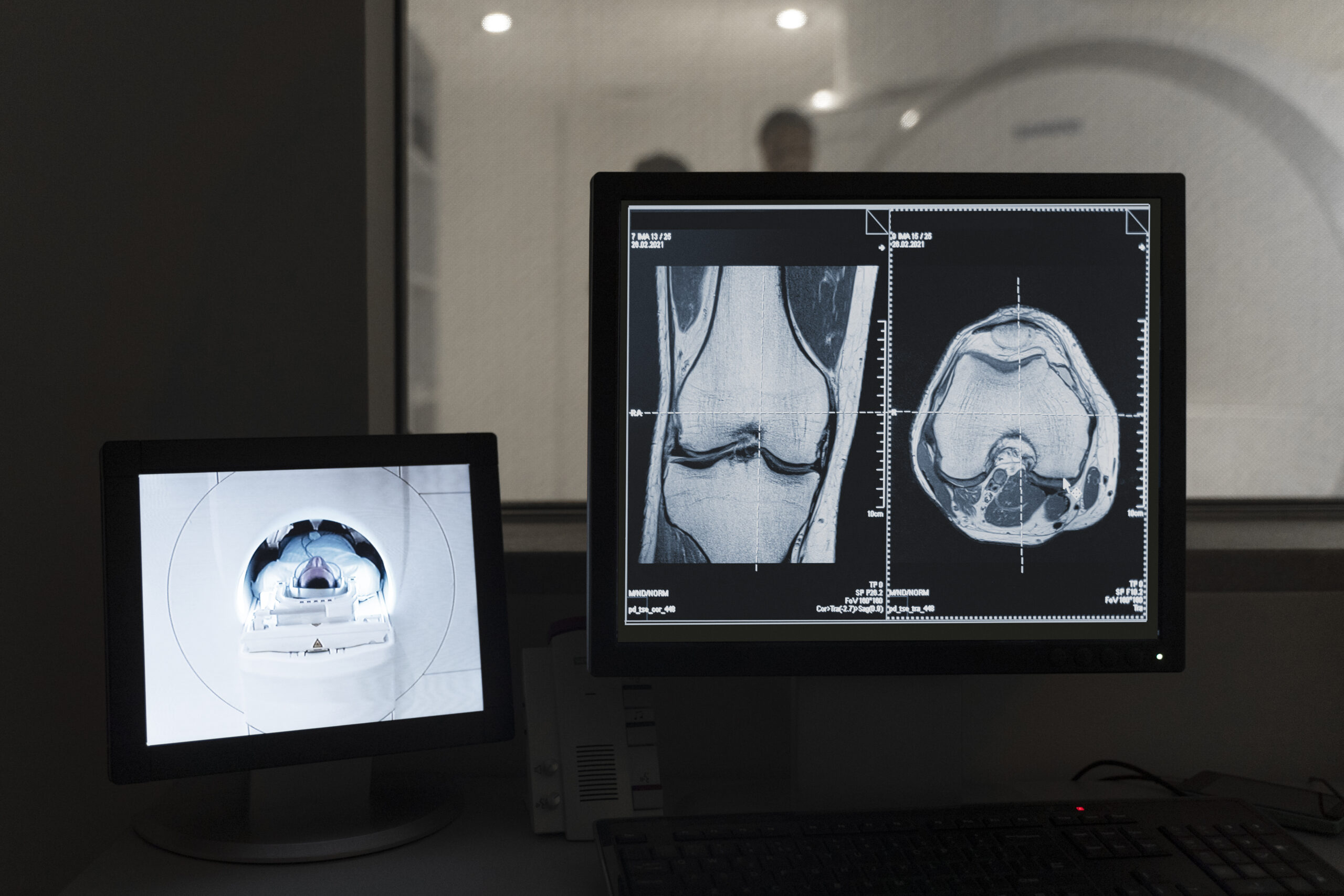
Medical scribes are trained to efficiently navigate electronic health records (EHR) systems. This proficiency results in quicker access to patient information, quicker note-taking, and faster turnaround times for lab orders and prescriptions. This increased efficiency translates into shorter wait times for patients and more focused, timely care. - Enhanced Patient-Physician Interaction:
With medical scribes handling documentation, physicians can devote more attention to their patients. They can engage in meaningful conversations, actively listen to patient concerns, and develop personalized care plans. This improved patient-physician interaction not only fosters trust but also allows for a better understanding of patient needs. - Reduction in Physician Burnout:
The administrative burden in healthcare has contributed to high levels of physician burnout. Medical scribing services alleviate this burden by reducing the time spent on paperwork. Physicians can leave work with a sense of accomplishment, leading to increased job satisfaction and reduced burnout rates. - More Accurate Billing and Coding:
Scribes ensure that documentation accurately reflects the care provided, leading to improved billing and coding accuracy. This can result in fewer claim denials, reduced financial stress on healthcare organizations, and better reimbursement rates, ultimately benefiting patients by preserving the viability of healthcare facilities. - Rapid Charting and Consultation Notes:
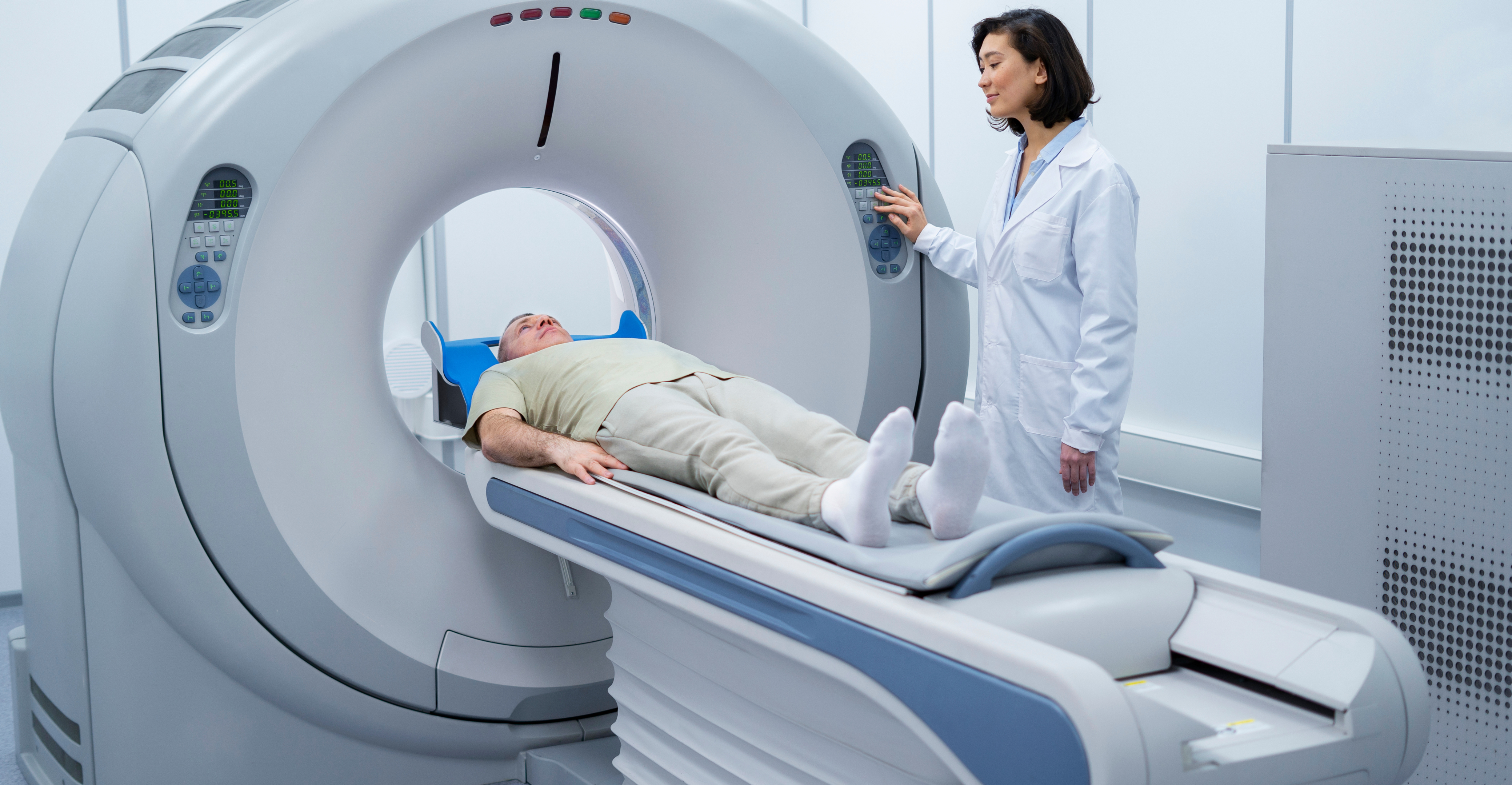
In fast-paced emergency departments and busy clinics, timely charting is crucial. Medical scribes can rapidly document notes during consultations, ensuring that critical information is readily available for follow-up care or referrals. - Comprehensive Clinical Support:
Medical scribes are trained in medical terminology and protocols, making them valuable clinical support members. They can assist with gathering patient histories, ordering tests, and facilitating communication between healthcare providers, further enhancing the quality of patient care. - Adaptability to Various Specialties:
Medical scribing services are adaptable and can cater to the needs of various medical specialties, from primary care to specialized fields like surgery, cardiology, and emergency medicine. This versatility ensures that patients across the healthcare spectrum benefit from improved care and physician satisfaction.
In the U.S. healthcare system, medical scribing services have emerged as a powerful tool for enhancing patient care and physician satisfaction. By delegating time-consuming administrative tasks to scribes, physicians can refocus on their core mission: delivering high-quality healthcare. The result is a win-win situation – improved patient outcomes and happier, more fulfilled healthcare professionals. As the healthcare landscape continues to evolve, medical scribing services are poised to play an even more significant role in shaping the future of healthcare delivery.
Hire a medical scribe from Saince so that you can improve your patients’ satisfaction and reduce physician stress!